Introduction
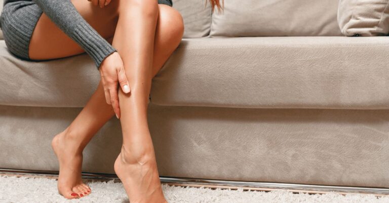
Finding a good reference for female leg drawing is not as difficult as it may sound. Here are some good places to look for one. The first place to start is a photograph. You can get a good picture of a woman’s leg from the internet. Alternatively, you can choose a photo of a male leg. You will need to take note of the details of the pose, such as the limb length and the head and neck.
Female leg drawing reference
When drawing the female leg, one of the most important aspects to consider is the proportions. The proportions of the leg are not necessarily the same as in reality, so it is helpful to look at photos of real people’s legs to get a better idea of what they look like. If you are unsure of the proportions of a leg, you can also use a video tutorial to teach you how to draw a leg.
To draw a female leg, you’ll need to know how to draw the foot and the legs. To start, sketch in the foot and then the first toe. Next, draw a “J” shape with the other toes. In step eight, shade the leg. Shade the lower leg and the front leg the most, with the thigh and calf getting lighter shaded. Follow these steps to create a beautiful female leg.
Female leg reference
You can find numerous female leg drawing reference images on the web. These can be used for a variety of purposes, including drawing a leg from a cartoon or an anime. Using real photos of the legs of your favorite character or actress is also helpful for drawing a female leg. You should also have some knowledge of leg proportions and shapes, because the proportions of legs in anime and manga tend to be identical to those of real legs.
You can also find various poses of the human female for drawing reference. It is important to remember that the human female has a heavy set body, so the leg of the right leg is bigger than the one on the left. These images can be referred to as anime female or girl poses. They can serve as good examples for beginners. Use blue lines as general structure guidelines, and red lines to indicate exposed skin. If you’re not confident with your drawing skills, you can always trace over the poses to get a general idea of the proportions.
Leg reference photo
First, you need to find a good leg drawing reference photo. There are many sites online that offer free stock photos, including Pexels and Unsplash. To get started, study many different photos of legs and notice how they curve. Once you have a few of these photos, you can practice drawing legs from different angles. Try creating a ball with three sets of legs. This ball can help you practice drawing legs from different angles.
Arm drawing reference
The first step in drawing an arm is understanding the proportions. Although the arm and head are both the same shape, people’s arm and head proportions will vary. This is mainly due to foreshortening in perspective. To understand this, it is helpful to study a model. Using a model can help you understand how to draw the arm proportionally. Then, you can try it yourself. If you find it difficult, you can always consult a reference sheet to draw it yourself.
Once you understand how the cylinders look, drawing an arm can be very easy. For example, the top sphere is the shoulder muscle, while the bottom sphere is the forearm. The forearm cylinder is thicker toward the elbow and tapered close to the wrist. A muscular person’s arm will be harder to draw, so you will need to exaggerate the muscles. Eventually, you will have to alternate between drawing reference and using your imagination to complete the arm.
Leg reference
Getting the right leg drawing reference is critical to creating a realistic illustration. You’ll need to understand the anatomy of the leg, which is one of the most difficult parts of the human body to draw. Having an accurate leg drawing will give your character a believable foundation and give you a starting point for your sketch. Taking a few moments to learn about leg anatomy and how it functions is a great way to enjoy the drawing process.
Draw a sketch of the leg to get an idea of its shape and movement. The next step in drawing a leg is to sketch the thigh and the lower leg. The thigh has a single bone while the lower leg is comprised of two bones, including the patella, a triangular bone that protects the knee joint. Then, shade the leg to show its contours. Once you have shaded and textured the basic shapes of the leg, add the details of toenails and the toe.
Foot reference drawing
If you want to learn how to draw feet, you should start by looking at a reference drawing. Observe the curve on the foot and compare it to the contours on your reference. Also, note the bony bits on the side of the foot. These are landmarks. Use the reference to mark them. The bones on either side of the foot represent the fibula and the base of the tibia, two parts of the leg.
Foots can be difficult to draw as they have multiple angles and forms. As long as you can break the foot into simple geometric shapes, you should be able to draw a good foot. As with any subject, practice makes perfect, so start practicing drawing feet today! Follow the steps below to draw great feet! Once you have mastered the basics, you can move on to more complex objects. For a more realistic representation, try using a digital reference drawing.
How to draw legs
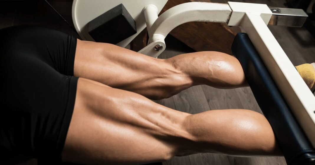
If you are learning how to draw legs, you’ll need to be aware of the differences between male and female leg shapes and sizes. Male legs are usually thicker and more muscular, with rounder calf muscles that create a dramatic taper from hip to ankle. On the other hand, women’s legs tend to be thinner and have more curves at the top of the thighs and hips. Drawing male and female legs is not difficult once you understand the basic anatomy of each.
Creating a leg drawing is easy once you understand how to imagine it in your mind. You can begin by drawing a straight line representing the lower leg and the hip. You can add details later. Then draw a notch at the bottom of the leg to represent the ankle. Once this is done, the rest of the leg can be drawn. The legs should have a distinctive color and texture. It’s also important to remember that drawing legs is a process of trial and error.

Torso drawing reference
When learning to draw the torso, it helps to have reference images of the torso to assist you. You should know the different plane directions to analyze your drawing problem. It is also helpful to have reference images of chests, as they help you create a clear understanding of the shape. Here are some ways to create a reference image of the torso. It’s not hard to draw a woman’s torso!
One of the most common stumbling blocks when drawing the torso is a habit of drawing straight lines. In fact, the torso articulates through leaning forward, back, and side-to-side. Try to avoid straight lines and perfect horizontal and vertical angles; small tilts are better and look more natural. Try drawing a woman who has a relaxed, slouchy appearance. Likewise, a person with a strong, muscular body is a great model to use as a reference for drawing the torso.
Leg Drawing Reference
Sketching the anatomy of the human leg is fun. There are so many things to study and learn about leg anatomy. In this article, you’ll learn the functions of the calf muscle and Banding lines. You’ll also learn about the femur, the long bone at the back of the thigh. By following these guidelines, you can create realistic-looking illustrations of the human leg. And don’t forget to enjoy the process!
Anatomy of the human leg
The major bones of the human leg are the femur, tibia, and fibula. There are also nerves, vessels, and bones that make up the leg. The leg can be examined easily using a good anatomy model. The most prominent bony prominences include the anterior superior iliac spine, the greater trochanter, and the medial malleolus. The anatomy of the human leg can be illustrated with a variety of tools, including a microscope or surgical instruments.
The lower leg is comprised of the lateral compartment, also known as the evertor compartment. The lateral compartment is bounded medially by the lateral surface of the fibula and laterally by the deep fascia of the leg. The fibular nerve, which originates on the superior two thirds of the lateral surface of the fibula, runs throughout the leg. A number of blood vessels supply and drain this area.
When sketching the leg, artists usually start by focusing on the athletic type because it has the most distinct muscle shapes. The legs of most people are generally cylindrical and feature few distinct muscular shapes. A typical skeletal drawing of a human leg will show the quadriceps and calf muscles, which are the most common muscles. The legs also include the patella, a triangular bone that protects the knee joint.
The lymphatic system in the legs is comprised of blind-ending lymphatic capillaries that drain fluid to the thoracic duct. The lymphatic system of the extremities consists of a network of subfascial collectors and perforating lymphatic veins. The epifascial lymphatic system in the human leg is divided into dorsolateral and ventromedial bundles.
Functions of the calf muscle
The calf muscle is a large flat muscle located in the lower leg. It joins with the soleus to form the Achilles tendon, which attaches to the heel bone. The calf muscle has two heads, the medial and lateral, and starts below the knee. Because it connects to two joints, it is prone to injury. In leg drawing reference, the medial head comes from the medial condyle of the femur, while the lateral head comes from the outside.
The quadriceps are composed of four individual muscles that attach at the knee. The quadriceps are shaped like a teardrop, while the calf is mainly defined by two calf muscles. These muscles come from the rear of the leg and extend into the heel. Other muscles and tendons make up the front and sides of the leg. The semitendinosus and the adductor longus are grouped together in the inner thigh, which is usually drawn as a triangle.
This study aimed to identify the risk factors for the common calf strain injury. The authors found that the best risk factors for this injury were not the type of exercise performed, but the time spent on activity. Athletes with CMSI were more likely to experience recurrent injuries and subsequent lower limb injuries. The authors found that preseason screening may not predict the severity of the injury, but it is helpful to know the risks so that prevention can be adapted to specific circumstances.
The calf is the most important muscle in the leg. It helps propel the leg forward and allows the knee to lock and rotate. It also plays a role in jumping and running. Among its functions are locking and rotating the knee. This makes the calf an important part of leg drawing reference. And when it is injured, the muscle should be treated right away. It may be painful.
Banding lines
If you are attempting to draw a leg, you should start by creating a full size image of the leg. Make sure to align the vertical axis down the center of the leg and the horizontal axis perpendicular to the center line. Make sure to mark the base of the apron, which is the bottom edge of the banding and the start of the taper. Also, be sure to mark the centerpoints of all arcs. This can be done by taking two parallel lines and finding their intersection.
Now, it’s time to draw the legs. To start, draw a straight line from the top to the bottom of the leg. It doesn’t matter if the leg is short or long. The bottom and top notch should represent the hip and ankle. Add details to these lines later. For the time being, do not worry about the banding lines. It’s all about understanding the anatomy of the leg so you can draw it accurately and creatively.
The banding lines run from the glutes to the back of the knee. They are also very helpful for filling in the inner thigh. The Biceps femoris is another muscle to fill out the inner thigh. This muscle flexes the knee. Begin with it in the thigh and extends to the head of the fibula near the knee. Then, follow this outline with the rest of your leg to complete the leg.
Anatomy of the femur
The femur is a long, cylindrical bone that is split into three major regions, the head, the body, and the shaft. The head is formed by a convex anterior surface and is separated from the body by the lateral and medial borders. The body is made up of two ridges that straddle the femur, the greater trochanter and the lesser trochanter.
The head of the femur is a roughly spherical structure that sits superomedially and projects anteriorly from the neck of the thigh. This smooth convexity is interrupted by a depression that is the fovea capitis femoris. To better understand the structure of this bone, consider using diagram labeling activities and quizzes to learn the various parts.
The superior femur articulates with the acetabulum in the pelvis. The superior femur articulates with the acetabulum, which forms the lateral portion of the femur. A femur that is properly positioned in the pelvis will help you walk more comfortably. Likewise, one that has an angular shape will make it easier to balance while walking.
The femur’s blood supply is vital for the health of the leg. Although most femur pathologies are traumatic, they can be caused by nontraumatic events, such as fractures. Examples of such nontraumatic events are a slipped capital femoral epiphysis and femoroacetabular impingement. There are also neoplastic conditions that can affect the femur.
The lateral ridge is a highly informative part of the femur, which varies considerably in size. Some species have poorly developed or missing lateral ridges. Others are characterized by a pronounced lateral ridge. The femur’s head shape depends on the species, but generally speaking, the femur is similar among phyllostomids and mormoopids.
Anatomy of the humerus
When you look at the anatomy of the humerus, you’ll notice that it’s composed of several different parts. The humerus’ head is the ball-shaped part, and it forms a ball-and-socket joint with the scapula. In addition, the head is large, forming about one-third of the entire sphere. It has a groove at the end, called the anatomical neck, where the shoulder capsule attaches.
The humerus is made up of two different layers, the articular portion, which is wide transversely and articulates with the radius and ulna, and the non-articular part, which has a pulley-like surface. The articular portion is comprised of the trochlea, which is a narrow, slender ridge, and a medial epicondyle.
The humerus is the largest bone in the upper arm and articulates with the scapula and ulna to form the elbow joint. The humerus is divided into the head and shaft region, and has two ridges called tubercles. These ridges provide stability and movement. They are important for preventing scapular injuries. Anatomy of the humerus consists of the articulation between the scapula and humerus, allowing the humerus to be a stable joint.
There are numerous anatomical features of the humerus, and a mnemonic can help you remember them. Anatomical features at the distal end are called cranial crest, scapula, and acromioclavia. These fractures occur most often in older adults with osteoporosis and are caused by an impact. During an impact, force from the fall on the hand often fractures the humerus.


